Dislocated Kneecaps in Pets
The knee is a complex structure consisting of muscles, ligaments, tendons, cartilage and bones. These components must align properly and interact harmoniously in order to function properly. Three bones are included in the knee: the femur, the tibia and the patella (kneecap).
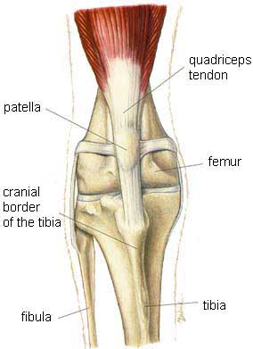
Normal Knee Joint of a Dog
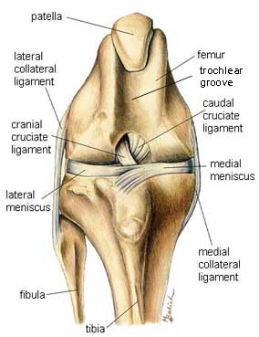
Normal Exposed Knee Joint of a Dog
Patellar luxation (kneecap dislocation) is a condition where the kneecap does not align properly with the femur and tibia. The condition can be temporary or permanent and range from complete dislocation to mild patellar instability. The dislocation can occur laterally (toward the outside of the knee joint), medially (toward the inside) or in both directions.
There are four types of patella luxations.
Grade 1: The patella is positioned normally but can be luxated with slight manual pressure.
Grade 2: Spontaneous luxation occurs; however, it can reduces spontaneously or can be replaced manually.
Grade 3: The patella is luxated most of the time; however it can be replaced manually.
Grade 4: The patella cannot be reduced manually.
Most commonly, the disease is a medial luxation and occurs as a result of a congenital (existing at birth) condition in toy and miniature breeds of dogs. Dogs with medial patellar luxation exhibit a bow-legged stance. In older animals, the condition appears suddenly and is often the result of a traumatic injury.
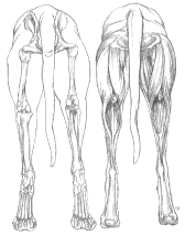
Stance of a Normal Dog
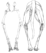
Bow-Legged Stance of Dog with Medial Patellar Luxation
Mild to severe lameness is the major clinical symptom. Non-weight bearing lameness and pain on manipulation of the knee joint are often associated with traumatic patellar luxation.
Patellar luxation is occasionally seen in cats. The condition is generally a medial luxation associated with a non-painful lameness. Surgery is recommended if the lameness persists.
Treatment for patellar luxation varies from one animal to the next. Animals that show no sign of pain or lameness are treated conservatively. This treatment includes anti-inflammatory medication, restricted exercise and rest. Animals that are affected more severely are generally candidates for surgery.
Below are photos taken of a dog who required surgery to correct a grade 4 patella luxation. Since most surgical corrections of patellar luxations consist of deepening the groove in which the patella rides, a trochlear wedge recession was performed on this dog. By deepening the groove in which the kneecap rides, this dog’s patella would be less likely to move into an abnormal position. This surgical technique is usually combined with other techniques in order to maximize stability of the knee.
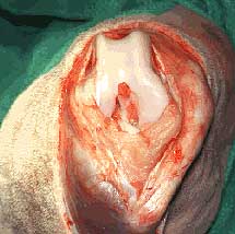
Exposed Trochlear Groove
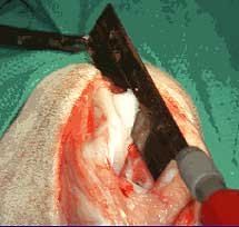
Begin Trochlear Wedge Recession
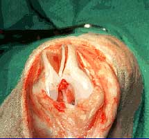
Continuation of Surgery
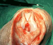
Trochlear Groove is Deepened and Ready for Patella
The objective of any surgery aimed to correct patella luxation is to stabilize the kneecap anatomically (in the trochlea) while maintaining full range of pain-free motion. The outcome following surgery is generally satisfactory; however, it depends on the individual care and severity of arthritis prior to surgery. Unfortunately, recurrence rates are frequent and luxations may either be in the same direction or other direction but are usually of lower grade than the original luxation.